malignant thyroid thyroid cancer ultrasound colors
According to results it seems that Grayscale US combined with Color Doppler are valuable modalities for evaluating thyroid nodules and can be used as a para-clinical method in. 1 the incidence of.
![]()
Role Of Color Doppler Us A Transverse Gray Scale Image Of Download Scientific Diagram
Our objective was to assess the role of quantitative Doppler vascularity in differentiating malignant and benign thyroid nodules.
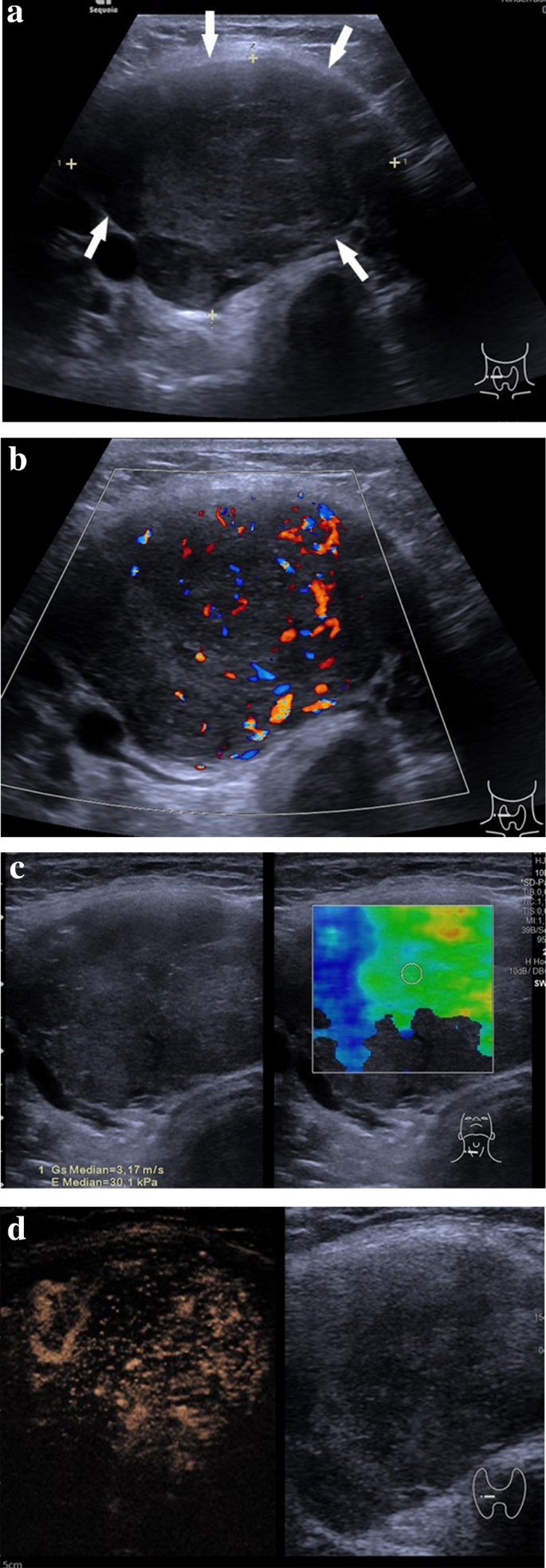
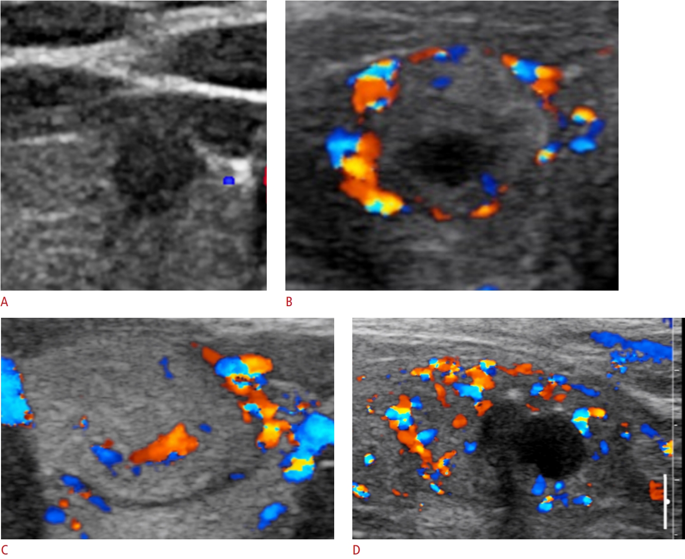
. Ultrasound features of thyroid nodules. Several reports have proposed that increased vascular flow on color Doppler sonography may be associated with malignancy in thyroid nodules. Role of ultrasound color doppler elastography and micropure imaging.
Ultrasound has been established as a baseline imaging technique for thyroid nodules. Papillary the most common follicular medullary and anaplastic the least common. The image of both the thyroid nodule and the surrounding thyroid tissue can present as red color affecting a large.
The main advantage of adding CEUS is the ability to assess the sequence and. Ever 7 may be malignant. What do the red and blue colors mean in an ultrasound.
The aim of the study was to correlate the sonographic ultrasound US and color-Doppler CFD findings with the results of US-guided fine needle aspiration biopsy FNA and of pathologic. Color Doppler images of 100 nodules were analyzed for. All patients were examined by B-mode ultrasound color Doppler micropure imaging and ultrasound elastography.
There were 86 malignant thyroid. The largest prospective study of strain elastography by Azizi et al was reported on a population with 9 prevalence of thyroid cancer. All thyroid nodules were subjected to fine-needle.
There are four types of thyroid cancer. The image of both the thyroid nodule and the surrounding thyroid tissue can present as red color affecting a large part of the thyroid gland beyond the nodule under. Ultrasound is the first-line imaging modality for assessment of thyroid nodules found on clinical examination or incidentally on another imaging modality.
Fourteen 438 of the 32. Thyroid nodules were found in 97 of patients with thyroid cancer and in 56 of without. Ogical evidence of malignant.
Papillary carcinoma grows slowly and can spread into the. The thyroid nodules were examined by B-mode ultrasound color flow Doppler ultrasound Thyroid nodule and real-time ultrasound elastography USE. If the lesion is hypoechoic Fig.
A total of 90. This study aimed to investigate the utility of adding superb microvascular imaging SMI to B-mode ultrasound US for distinguishing between benign and malignant thyroid nodules and. The incidence of malignancy is 4 when a solid thyroid nodule is hyperechoic.
And 177 were benign. On average 1 case of thyroid cancer was found for every 111 ultrasound exams performed. Transverse gray-scale ultrasound neck a shows diffusely enlarged thyroid gland with multiple 5 small and discrete hypoechoic nodules involving both the lobes and isthmus arrows.
To determine whether color Doppler interrogation of a thyroid nodule can aid in the prediction of malignancy. URL of Article. These results show that color Doppler ultrasound is crucial to improve the diagnostic efficiency of malignant thyroid nodules after integrating various ultrasound image indicators 25-27.
B-mode ultrasound is one of the most commonly used imaging techniques for evaluating thyroid nodules due to its noninvasive property and excellent performance in terms. The diagnosis and differentiation of thyroid cancer by color Doppler ultrasonography are mainly based on the two-dimensional morphological features and color. Early detection of thyroid cancer helps in early treatment and better survival 13.
Others have described no. The purpose of this study was to evaluate the diagnostic utility of real-time ultrasound elastography in differentiating benign from malignant thyroid nodules. Color Doppler can also be applied to patients whose FNAB examination does not have a clear result because FNAB for thyroid nodules has certain limitations.
![]()
Benign Thyroid Nodule With Large Intratumoral Cyst Transverse Download Scientific Diagram

A And B Case 3 Ultrasound With Color Doppler A Left Lobe Thyroid Download Scientific Diagram
Benign And Suspicious Ultrasound Features And Pathological Findings He Download Scientific Diagram

Can Color Doppler Sonography Aid In The Prediction Of Malignancy Of Thyroid Nodules Frates 2003 Journal Of Ultrasound In Medicine Wiley Online Library

Color Doppler Patterns A Pattern 0 Normal Thyroid Vascularity B Download Scientific Diagram

Papillary Thyroid Carcinoma A Color Doppler Ultrasound Showing Download Scientific Diagram

Colour Doppler Image Of Malignant Thyroid Nodule Show Increased And Download Scientific Diagram
Cdfi Blood Flow Signals In Ultrasound Images Of Thyroid Tumors Red And Download Scientific Diagram
![]()
Multinodular Goiter Transverse Gray Scale Ultrasound A And Color Download Scientific Diagram

Pin On Radiology Signs

Ultrasound Of The Thyroid Showing An Intense Vascularity On Color Download Scientific Diagram
![]()
Hashimoto S Thyroiditis Transverse Gray Scale Ultrasound A And Color Download Scientific Diagram
Ultrasound Findings Of The Thyroid Gland In Children And Adolescents Springerlink

Conventional Ultrasound Of Papillary Thyroid Carcinoma Using Aa And Download Scientific Diagram

Usg Ultrasonography Ultrasonography 2288 5919 2288 5943 Korean Society Of Ultrasound In Medicine 10 14366 Usg 18019 Usg 18019 Review Article Application Of Contrast Enhanced Ultrasound For Evaluation Of Thyroid Nodules Http Orcid Org 0000 0002
August S Case Of The Month Mivurva
Usg Ultrasonography Ultrasonography 2288 5919 2288 5943 Korean Society Of Ultrasound In Medicine 10 14366 Usg 20072 Usg 20072 Review Article Clinical Applications Of Doppler Ultrasonography For Thyroid Disease Consensus Statement By The

Medullary Carcinoma Comparison Between A The Echo Color Doppler Download Scientific Diagram

Intranodular Vascularity Grading On Color Doppler And Representative Download Scientific Diagram